Regular hair and scalp assessments deliver five concrete advantages: earlier detection of alopecia subtypes and scalp disease, a chronological biochemical record that blood tests can't match, objective before-and-after treatment tracking, identification of suspicious lesions before they progress, and the psychological clarity that comes from knowing what's actually happening on your scalp.
The top five advantages at a glance:
- Detect alopecia subtypes, infections, and scalp disease earlier
- Use hair as a month-by-month biochemical archive (1 cm ≈ 1 month of data)
- Track treatment response with objective imaging, not guesswork
- Catch suspicious scalp lesions that routine medical visits routinely miss
- Reduce anxiety and improve treatment adherence with documented progress
The sections below explain each advantage with clinical evidence, cover the main assessment methods and their real limits, and tell you exactly who should get checked and how often.
Key Takeaways
Regular hair and scalp assessments deliver measurable diagnostic and monitoring advantages by combining hair's unique chronological record with objective imaging and clinical examination.
| Point | Details |
|---|---|
| Hair as a time capsule | Each centimeter of hair shaft stores about one month of biochemical data, enabling retrospective diagnosis. |
| Early lesion detection | Checks every 8–12 weeks catch suspicious scalp lesions that annual medical visits routinely miss. |
| Objective treatment tracking | Trichoscopy and standardized photography remove recall bias and validate whether therapy is working. |
| Know the limits | Contamination, lack of standardized reference ranges, and averaging effects mean hair analysis needs clinical correlation. |
| Myhair for frequent monitoring | Myhair's AI scanner and scoring system make consistent, home-based photo tracking practical between clinic visits. |
Table of Contents
- What does a regular hair assessment actually include?
- What are the advantages of regular hair assessment for diagnosis?
- How do regular assessments improve treatment monitoring?
- Which assessment methods are worth knowing about?
- Who should get regular assessments, and how often?
- What are the real limits of hair assessment?
- How should you prepare for an assessment, and what happens next?
- How does AI-powered monitoring fit into regular hair assessments?
- Why frequent checks matter more than most people realize
- Myhair makes frequent monitoring practical
- Sources
- FAQ
What does a regular hair assessment actually include?
"Regular hair assessment" covers a spectrum of services, from a 10-minute visual check at a salon to a full trichologist workup. Knowing what falls inside that spectrum helps you ask for the right thing.
Typical components:
- Visual examination: Parting the hair in multiple locations to assess density, texture, and scalp condition
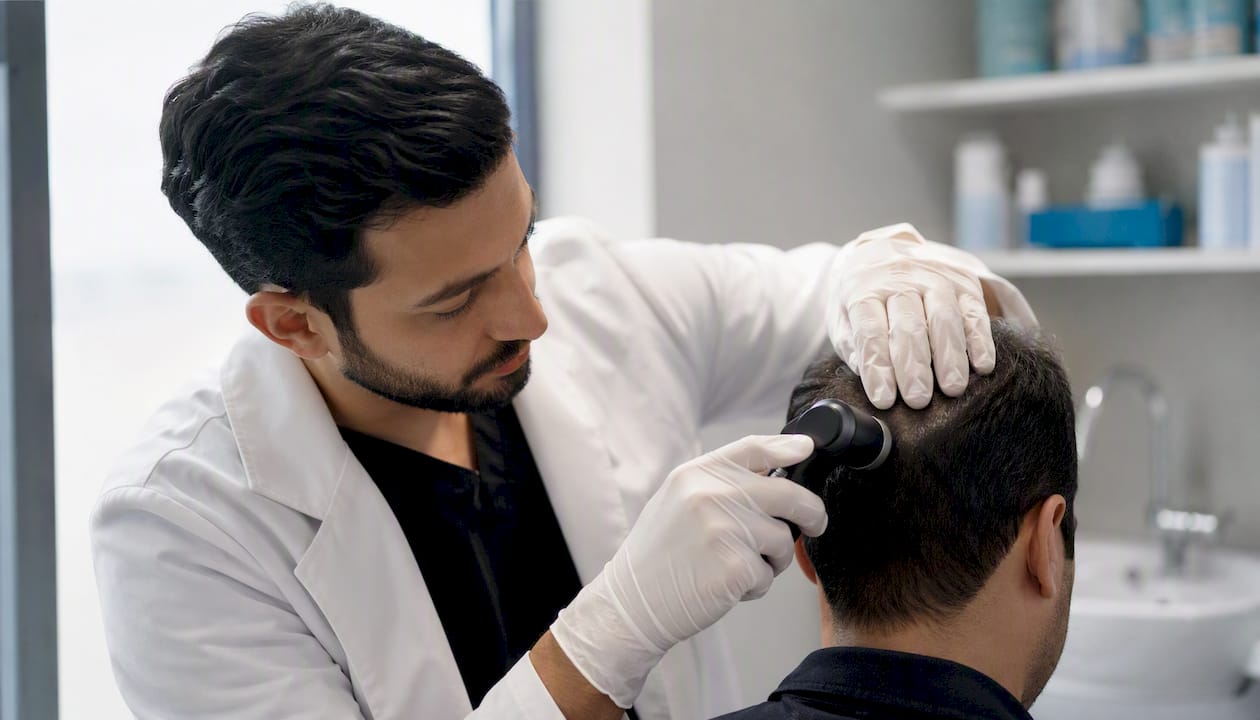
- Dermoscopy/trichoscopy: A handheld or video dermatoscope magnifies the scalp 10–70x to reveal follicle structure, vascular patterns, and scaling invisible to the naked eye
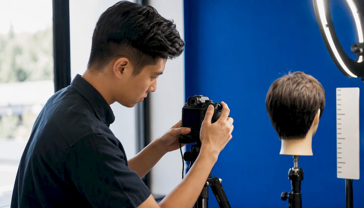
- Global photography: Standardized photos from fixed angles (top, crown, temples) to document density changes over time
- Gentle hair-pull test: Grasping 40–60 hairs and applying gentle traction; normally 0–2 telogen hairs are extracted, so extracting anagen hairs signals active pathology
- Wash/daily count: Counting shed hairs over a defined period to quantify loss rate
- Hair sampling (trichogram or biospecimen): Plucking or cutting hair for microscopic or chemical analysis
- AI imaging/app scans: Photo-based automated scoring that tracks density trends between clinic visits
"Regular" means different things depending on your goal. Cosmetic monitoring typically runs every 8–12 weeks. Medical monitoring during active treatment often means every 1–3 months. Toxicological or longitudinal biomarker tracking uses monthly hair segments because each centimeter of shaft represents roughly one month of physiology.
What are the advantages of regular hair assessment for diagnosis?
Hair is one of the few tissues in the body that functions as a passive recorder. Each centimeter of hair shaft stores roughly one month of biochemical data, meaning a 6 cm sample taken today carries a six-month retrospective window. Blood and urine give you a snapshot of right now; hair gives you the film reel.
That longitudinal record has direct clinical value across several conditions:
- Alopecia subtypes: Trichoscopy distinguishes androgenetic alopecia (miniaturized follicles, peripilar signs) from telogen effluvium (uniform shedding, no miniaturization) and alopecia areata (exclamation-mark hairs, yellow dots), guiding treatment before months of trial-and-error
- Scalp infections and inflammation: Dermoscopic patterns identify fungal infections (tinea capitis), seborrheic dermatitis, and psoriasis with enough specificity to guide topical therapy
- Suspicious lesions and skin cancer: Seeing clients every 8–12 weeks, hair professionals and trichologists can spot evolving moles or new scaling that patients never notice in a mirror. Cases where stylists identified scalp melanoma early have been documented in clinical reporting, and the scalp is a notoriously missed site at annual dermatology visits
- Trace-element and toxin exposure: Hair elemental analysis can screen for environmental and occupational exposures including heavy metals, though interpretation requires careful contamination controls
- Medication adherence and cortisol: Hair metabolites reflect cumulative drug intake and chronic stress hormones over weeks to months, a window no blood draw can open
Distinguishing chronic from acute shedding is one of the most practically useful diagnostic applications. A patient who presents with diffuse thinning today may have experienced a triggering illness or surgery three to four months ago. Regular assessments create the timeline that connects those dots.
How do regular assessments improve treatment monitoring?
Objective documentation is what separates a well-managed hair condition from an expensive guessing game. Without baseline images and periodic measurements, neither the clinician nor the patient can reliably tell whether a treatment is working, plateauing, or failing.
What structured monitoring actually delivers:
- Before/after imaging: Standardized global photos and trichoscopy scores taken at fixed intervals give a visual record that removes recall bias. Trichoscopy is validated for monitoring therapeutic response across common scalp conditions, including androgenetic alopecia and alopecia areata
- Dose optimization: Hair cortisol and medication metabolites measured in sequential segments allow clinicians to verify that a patient is actually absorbing and using a drug at therapeutic levels, not just reporting compliance
- Earlier course correction: A three-month photo review showing no density change is actionable data. It tells you to adjust the product, the dose, or the diagnosis before another three months pass
- Behavioral reinforcement: Seeing documented improvement, even modest, motivates continued adherence. Conversely, seeing no change early prevents patients from spending months on a product that isn't working for their biology
A practical three-month review workflow looks like this: standardized crown and temple photos at baseline, a trichoscopy session at week 12, comparison scoring, and a revised plan. That cycle, repeated consistently, builds a longitudinal record that hair's role as a biospecimen makes uniquely informative.
Pro Tip: Ask your provider to photograph from the same angle and lighting at every visit. Inconsistent photography is the single biggest reason before/after comparisons fail to show real change.
Which assessment methods are worth knowing about?
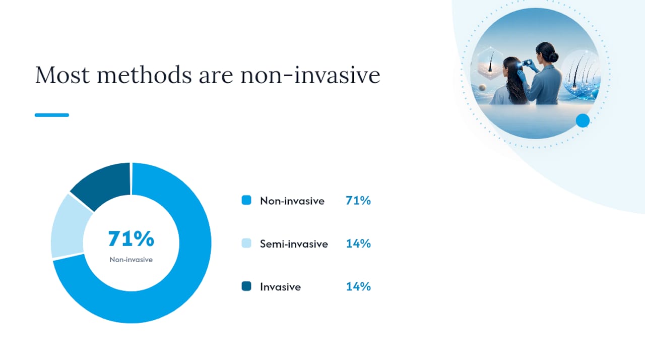
Clinical literature groups hair-evaluation methods into non-invasive, semi-invasive, and invasive categories, and no single method covers every diagnostic question. Here's how they compare in practice:
| Method | Invasiveness | Repeatability | Best use case | Main limits |
|---|---|---|---|---|
| Global photography | None | High | Density trend tracking | Requires standardized setup; misses microscopic changes |
| Trichoscopy/dermoscopy | None | High | Follicle pattern, scalp disease | Operator skill-dependent; no biochemical data |
| Wash/daily hair count | None | Moderate | Quantifying shed rate | Tedious; affected by washing frequency |
| Phototrichogram | None | High | Hair density and growth rate | Requires shaving a small area; time-consuming |
| Trichogram (pluck) | Semi-invasive | Moderate | Anagen/telogen ratio | Uncomfortable; sampling variability |
| Scalp biopsy | Invasive | Low | Definitive histological diagnosis | Scarring risk; single time point |
| Hair biospecimen (cut) | None | High | Toxicology, cortisol, metabolites | Contamination risk; no standardized reference ranges for many analytes |
A few things the table doesn't capture: trichoscopy's value scales with the operator's experience, and AI-assisted image scoring is beginning to reduce that variability by automating follicle counts and density measurements. Current AI tools add real value for frequent monitoring between clinic visits, but they depend on consistent image quality and still require clinical correlation for any diagnosis. They don't replace biopsy or specialized lab panels.
Who should get regular assessments, and how often?
Most people benefit from at least a periodic scalp check, but certain groups have a stronger clinical case for structured, frequent monitoring.
Higher-risk indicators that warrant regular assessment:
- Rapid or unexplained shedding (more than usual over several weeks)
- Family history of early-onset pattern baldness
- Autoimmune conditions (lupus, thyroid disease, alopecia areata history)
- Current or recent chemotherapy or immunosuppressive therapy
- Visible scalp lesions, new moles, or changing pigmentation
- Occupational exposure to heavy metals, solvents, or pesticides
- Recent major illness, surgery, or prolonged high stress (hair loss often lags the trigger by months)
Recommended cadence by scenario:
- Routine cosmetic monitoring: every 8–12 weeks, aligning with typical salon visits
- Active medical treatment (minoxidil, finasteride, PRP, etc.): every 1–3 months to assess response and adjust
- Toxicological or biomarker tracking: monthly hair segments, since each centimeter covers approximately one month of exposure history
Finasteride is FDA-indicated for men only. Women who are or may become pregnant must not handle crushed or broken finasteride tablets, due to the risk of harm to a male fetus. It can also cause sexual side effects (reduced libido, erectile dysfunction) in a minority of men, which usually resolve after stopping treatment. Discuss these risks with a physician before starting.
That growth rate is also why timing matters for sampling. A sample taken from the root captures the most recent month; the same strand's distal end may reflect events from a year ago.
What are the real limits of hair assessment?
Hair analysis is genuinely useful, but it has well-documented limitations that any honest practitioner will tell you upfront.
Key limitations:
- External contamination: Shampoos, dyes, bleach, and environmental deposits can alter elemental readings. A single chemical treatment can make a hair mineral analysis unreliable without careful controls
- No standardized reference ranges: For many analytes, there are no universally agreed normal ranges, so a "high" result from one lab may be unremarkable at another
- Averaging effect: Hair captures cumulative exposure, not acute events. A single toxic exposure that resolved quickly may not register clearly in a monthly segment
- Sensitivity limits: Hair tests are not sensitive enough to detect all acute or low-level exposures, and a normal result does not rule out pathology
Common pitfalls to avoid:
- Over-interpreting a single measurement without a trend line
- Relying on a vendor's proprietary score without clinical correlation
- Ignoring the effect of recent dyeing or bleaching on elemental results
- Treating a hair biomarker result as a standalone diagnosis
When to escalate beyond hair assessment:
Red flags that require blood tests, endocrine panels, or scalp biopsy include: sudden diffuse shedding with fatigue or weight change (thyroid or iron panel), scarring alopecia patterns on trichoscopy (biopsy needed for definitive diagnosis), a suspicious pigmented lesion (immediate dermatology referral), or any hair loss that fails to respond after 6 months of documented treatment.
Pro Tip: When requesting a referral, phrase it specifically: "I have documented progressive thinning over six months with trichoscopy images showing follicle miniaturization — I'd like an endocrine panel and a dermatology consult." Specific language gets faster specialist access.
How should you prepare for an assessment, and what happens next?
A little preparation makes the difference between a useful assessment and one that produces ambiguous results.
Pre-visit checklist:
- Avoid heavy oils, dry shampoo, or styling products for 24–48 hours before the appointment
- Skip chemical treatments (color, relaxers, perms) for at least two weeks prior if a hair sample will be collected
- Write down all current medications, supplements, and any recent illnesses or surgeries, including approximate dates
- Note any significant stressors from the past 3–6 months, since hair loss often reflects events that happened months earlier
- Bring previous assessment photos or reports if you have them
During the assessment:
The provider will typically part the hair in several locations, examine the scalp under dermoscopy, take standardized photos, and may perform a gentle pull test. If a biospecimen is needed, a small bundle of hairs is cut close to the scalp (not plucked, for most chemical analyses). The whole process usually takes 20–45 minutes for a thorough workup.
After the assessment:
Expect a written report or image set within a few days for in-person visits. Useful improvement in density or follicle health typically takes 3–6 months to appear in photos, so a single follow-up image at week four tells you almost nothing.
How does AI-powered monitoring fit into regular hair assessments?
AI-assisted hair analysis occupies a specific and genuinely useful niche: frequent, standardized image-based monitoring between clinic visits. It doesn't replace trichoscopy, biopsy, or lab panels, but it fills a gap that traditional clinical care leaves open.
Myhair uses a scanner camera and AI scoring algorithms to analyze uploaded hair scans, generating a hair score that tracks density and pattern trends over time. The practical value is in the frequency: most people see a dermatologist or trichologist once or twice a year, but meaningful hair changes can develop over weeks. Regular app-based scans create a data trail that makes those annual visits far more productive.
Where AI monitoring adds real value:
- Frequent photo tracking that catches pattern changes between clinic appointments
- Automated scoring that removes the subjectivity of self-assessment
- Trend visualization that helps users and clinicians see whether a treatment is moving in the right direction
- Personalized product recommendations based on individual hair analysis insights
Honest limitations:
- AI scoring depends entirely on image quality and consistent lighting; a blurry or poorly lit scan produces unreliable output
- The platform does not diagnose medical conditions and cannot replace clinical dermoscopy for identifying follicle-level pathology
- Suspicious lesions, scarring patterns, or rapid unexplained shedding require in-person clinical evaluation regardless of what an app score shows
Clinicians interested in integrating AI-assisted monitoring into practice can find technical and research information on the Myhair site.
Why frequent checks matter more than most people realize
There's a tendency to treat hair loss as something you address once it becomes obvious, and that instinct is exactly backwards. The conditions that respond best to treatment, androgenetic alopecia, early telogen effluvium, scalp inflammation, are the ones caught before significant follicle miniaturization or scarring occurs. By the time hair loss is visually dramatic, the treatment window for full recovery has often already narrowed.
What regular assessments do is shift the timeline. A person who checks in every 8–12 weeks with standardized photos and a trichoscopy session has a documented baseline. They know what "normal" looks like for their scalp. When something changes, they catch it at the two-month mark instead of the two-year mark.
The psychological dimension is real too, and it's underappreciated in clinical discussions. Anxiety about hair loss is often worse than the loss itself, partly because of uncertainty. A structured assessment replaces that uncertainty with data. Even a result that confirms ongoing thinning is more manageable than not knowing, because it comes with a plan.
For someone with early diffuse thinning and no clear diagnosis, the routine I'd suggest: a baseline trichoscopy with global photos, a blood panel ruling out thyroid and iron issues, and a three-month photo check with a consistent AI scan in between. That combination covers the clinical bases without over-medicalizing a situation that may resolve on its own.
Myhair makes frequent monitoring practical
Most people won't book a trichologist every eight weeks. The cost and logistics make consistent clinical monitoring unrealistic for the majority of people who would genuinely benefit from it. Myhair addresses that gap directly: upload a scan, get an AI-generated hair score, and track your trend over time from home.
The Myhair scanner camera captures standardized images that feed into the platform's scoring algorithm, giving you a repeatable baseline that makes every future scan comparable. That consistency is what turns a series of photos into actual trend data. For people managing androgenetic alopecia, recovering from telogen effluvium, or simply wanting to know whether a new product is doing anything, that trend line is the whole point.
Myhair does not replace clinical care for suspicious lesions, scarring alopecia, or any condition requiring biopsy. If your assessment flags something that needs a dermatologist, the platform's output gives you documented evidence to bring to that appointment. Start with the app onboarding to set up your first scan and baseline score.
Sources
- Human hair as a diagnostic tool in medicine - PMC
- Principles of dermatological practice. Examination of hair and scalp
- How your hairdresser could save your life | Wellcome Collection
- Trace element hair analysis and its diagnostic applications (review) - PubMed
- Hair evaluation methods: merits and demerits - PMC
- Trichoscopy in clinical evaluation and follow-up of hair and scalp disorders (Dermatologic Therapy)
This article is general information, not a substitute for advice from a qualified doctor. Consult a qualified healthcare professional about your own circumstances before acting on anything here.
FAQ
What are the main benefits of hair analysis testing?
Hair analysis can detect alopecia subtypes, trace-element exposures, medication metabolites, and chronic stress hormones across a retrospective window of weeks to months, something a single blood draw cannot do. Its primary limitation is that results require careful contamination controls and clinical correlation before any diagnosis is made.
Is a scalp analysis worth it?
For anyone with unexplained shedding, a family history of hair loss, or an active scalp condition, a structured scalp analysis with trichoscopy and standardized photography provides objective data that significantly improves treatment decisions. For routine cosmetic monitoring, even periodic checks every 8–12 weeks can catch changes early enough to act on them.
What can hair analysis help determine?
Hair analysis can help identify alopecia subtype and severity, scalp infections or inflammatory conditions, suspicious lesions, cumulative exposure to heavy metals or toxins, medication adherence, and chronic cortisol levels. The diagnostic value depends on which method is used and whether findings are interpreted alongside a clinical examination.
How often should you assess your hair?
Routine cosmetic monitoring works well on an 8–12 week cycle. Active medical treatment for hair loss typically calls for reassessment every 1–3 months. Toxicological or biomarker tracking uses monthly hair segments, since each centimeter of shaft covers approximately one month of exposure history.
Can Myhair replace a dermatologist for hair assessment?
Myhair's AI-powered scanning and scoring is designed for frequent photo-based monitoring and trend tracking between clinical visits, not for diagnosing medical conditions. Suspicious lesions, scarring alopecia patterns, or any rapid unexplained shedding require in-person clinical evaluation regardless of app results.